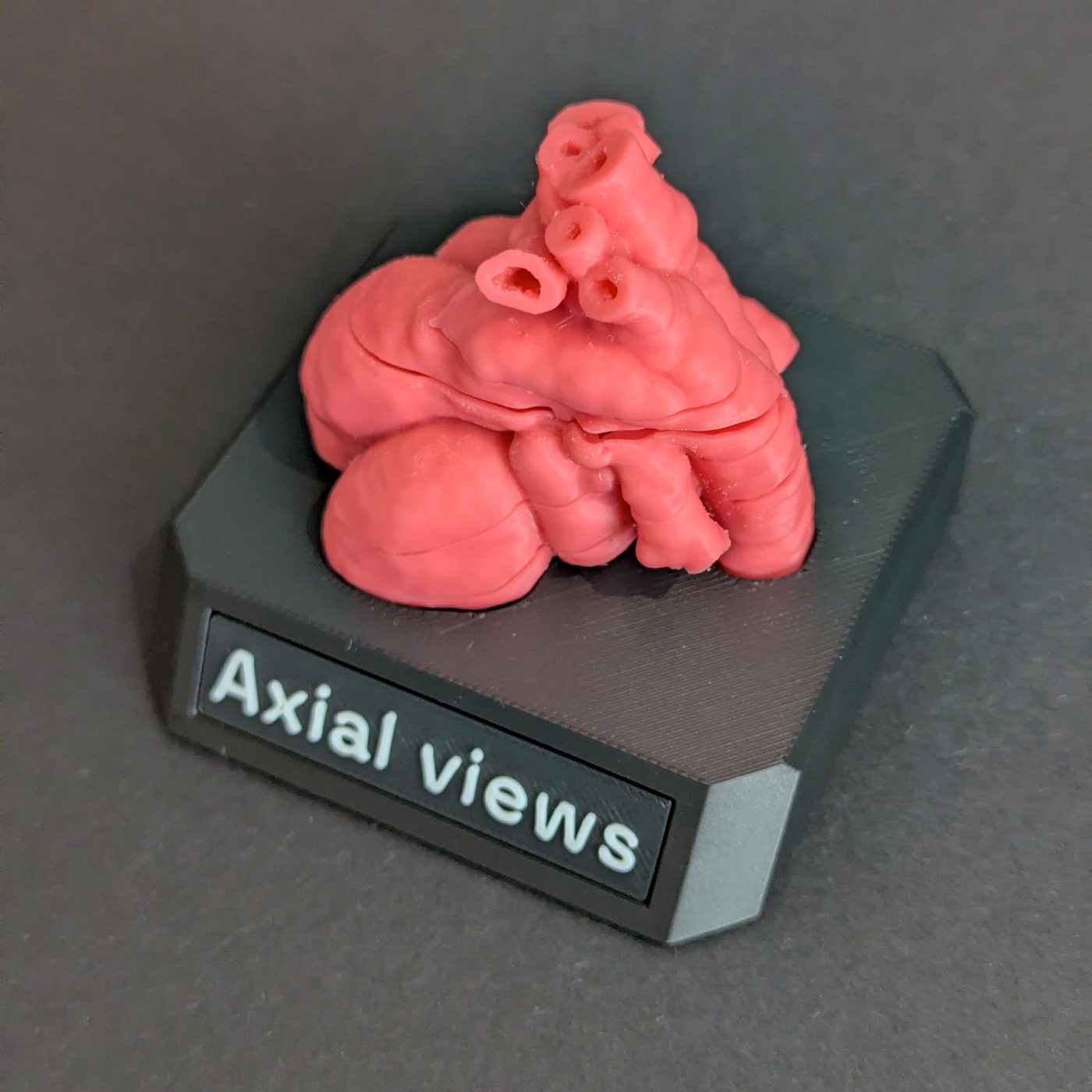
Fetal heart – Axial views, 1×
Four-chamber · Left ventricular outflow tract · Right ventricular outflow tract / three-vessel · Three-vessel and trachea
A life-size fetal heart in five magnet-located sections, cut along the four planes of the screening sweep.
Also available: 2× size.
New release price. Save £50 off the £149 list price until 9 January 2027, or until this first batch sells out if that is sooner.
Secure checkout by Stripe. Card details never touch our servers. By buying, you agree to our terms of sale.
About this model
The model is cut along the four diagnostic planes of the screening sweep, so each face you uncover is a view you already know how to read: the four-chamber view, the left ventricular outflow tract, the right ventricular outflow tract and three-vessel view, and the three-vessel and trachea view.
The heart comes apart into five sections, held together by magnets and stacked in the order of the sweep, from the four-chamber view at the bottom to the three-vessel and trachea view at the top. Lift them apart to see each view, then press them back together to put every view back in the heart it came from.
It comes with a stand, if you want to keep it on a shelf or a desk, and a velvet bag for when you take it with you. In the box too are four A6 cards, one for each diagnostic plane, each showing that plane's cut face with its structures labelled.
What it shows
The four cut faces are the four-chamber view, the left ventricular outflow tract, the right ventricular outflow tract and three-vessel view, and the three-vessel and trachea view.
Assembled, the sections also show how close together the views are. From the four-chamber plane to the three-vessel and trachea plane is about 17 mm through the middle of the heart, and the four-chamber and left ventricular outflow tract planes are about 7 mm apart.
At this scale it is a one-to-one or small-group object. For teaching a room, the 2× is the one to reach for.
Where the anatomy comes from
The geometry is the normal fetal heart atlas published by King's College London (Uus and colleagues, Journal of Cardiovascular Magnetic Resonance, 2022). The atlas is not a scan of one fetus. It combines the MRI of 28 healthy fetuses at 29 to 34 weeks: each scan was reconstructed in 3D with motion correction, all 28 were brought to a common size and averaged into one heart, and that heart was segmented into its chambers and vessels at 0.6 mm. Its cardiac circumference, measured on the four-chamber view, is that of a 34-week heart, and the 1× model is printed at exactly that size.
The atlas outlines the spaces inside the heart, the blood vessels and the trachea, not their walls. We build the walls around those spaces, with a thickness set for each structure, and then add the soft tissue the atlas does not contain: the mitral and tricuspid valves, the aortic and pulmonary valves, the moderator band, and the flap of the foramen ovale.
Specification
- Material
- PLA
- Source data
- SVRTK fetal MRI atlas, 0.6 mm isotropic
- Gestational age
- 29 to 34 weeks, third trimester
- Scale
- 1× at 34 weeks, about 54 × 41 × 52 mm (W × D × H)
- Pieces
- 5, each joint located by a raised lip and held by a magnet
- Planes shown
- Four-chamber · Left ventricular outflow tract · Right ventricular outflow tract / three-vessel · Three-vessel and trachea
See it unboxed and taken apart
Educational model, not a medical device. This is a teaching aid. It is not intended or approved for clinical diagnosis, treatment, surgical planning, or any patient-specific decision. The geometry comes from a published, anonymised fetal MRI atlas, not from an identifiable patient, and it represents atlas-average anatomy rather than any individual.