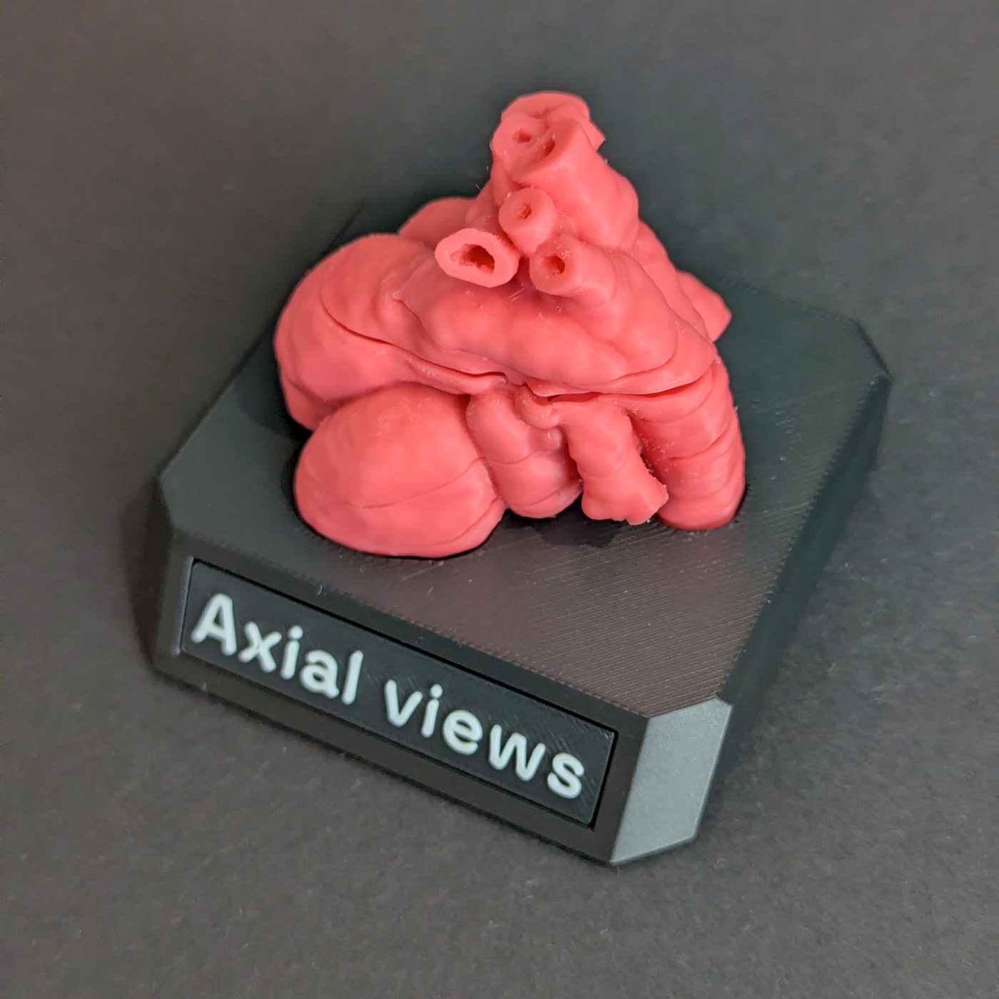
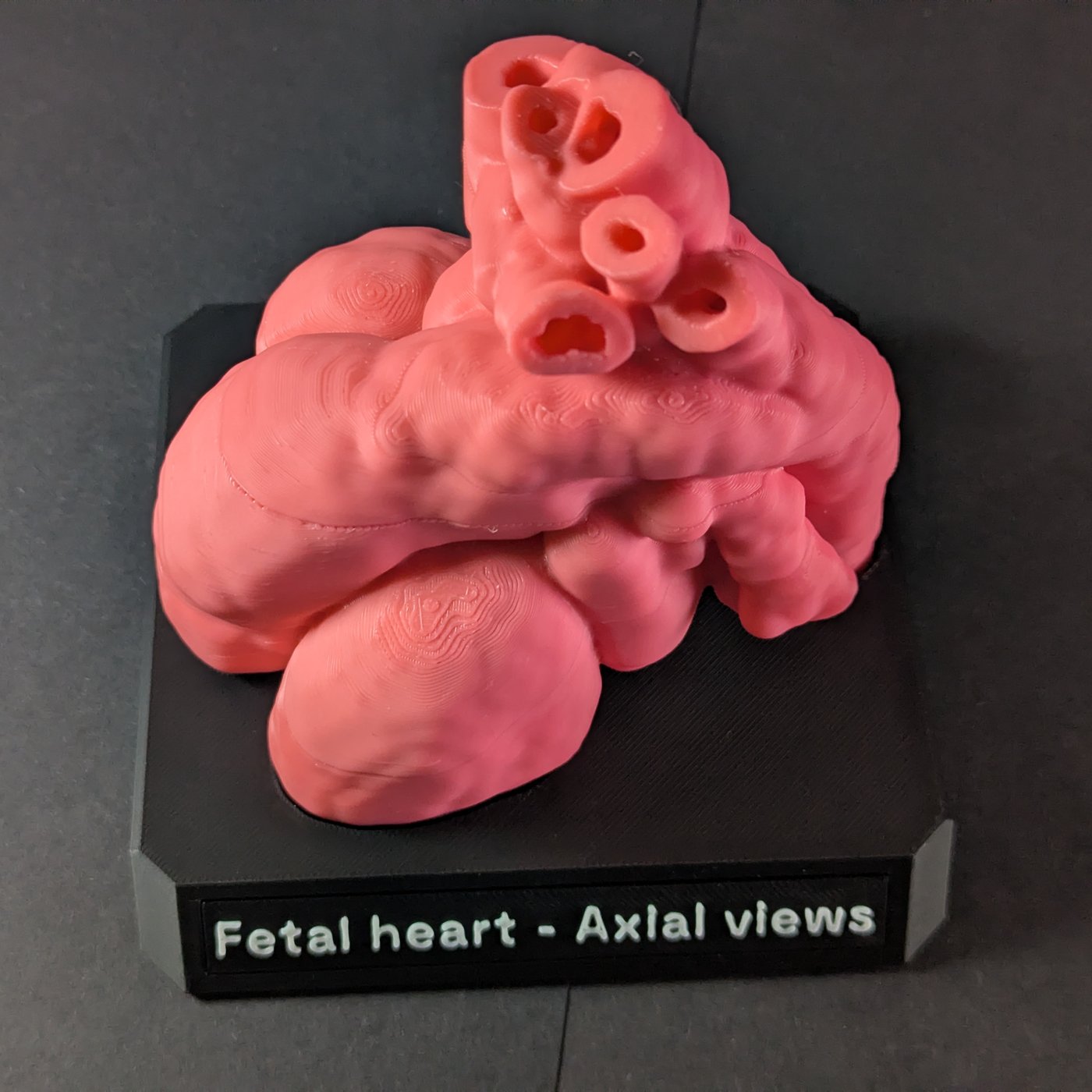
The four-chamber view, the left ventricular outflow tract (LVOT) view, the right ventricular outflow tract and three-vessel view (RVOT/3VV), and the three-vessel and trachea view (3VT) are the transverse, or axial, views of the fetal heart. They are taken in order as the probe moves up from the upper abdomen towards the fetal head, and all four lie within a short distance of each other. Below is what each view shows, what you check in it, and how the four fit together inside one heart.
The screening sweep
The ISUOG practice guidelines for fetal cardiac screening, updated in 2023, describe five transverse views taken in order as the probe moves cephalad. The first, across the upper abdomen, establishes situs. The other four are views of the heart, and are the ones covered here.
Each is a cross-section of the fetal chest at a different level, and often a slightly different angle. Fetal echocardiography adds further views, such as sagittal views of the aortic and ductal arches, but the transverse sweep is the core of the screen.
Four-chamber view
The first heart view on the sweep crosses the chest at the level of the atrioventricular valves and shows both atria and both ventricles at once.
What you check:
- the heart sits mainly in the left chest, takes up about a third of the thoracic area, and its axis points about 45 degrees to the left
- the two atria are of similar size, and so are the two ventricles
- the tricuspid valve inserts slightly closer to the apex than the mitral valve
- the moderator band sits near the apex of the right ventricle, which marks it as the morphological right ventricle
- the flap of the foramen ovale moves within the left atrium
- the pulmonary veins enter the left atrium
- the ventricular septum is intact from the apex to the crux
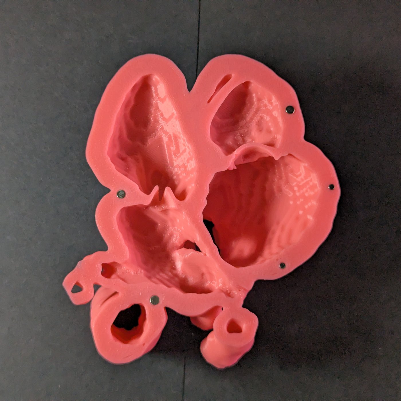
Left ventricular outflow tract (LVOT) view
Slide or tilt the probe slightly towards the fetal head from the four-chamber view and the aorta comes into view, leaving the left ventricle: that is the LVOT view.
What you check:
- the aorta arises from the left ventricle
- the anterior wall of the aorta runs on continuously from the ventricular septum
- the posterior wall of the aorta is continuous with the anterior leaflet of the mitral valve
- the aortic valve opens freely and is not thickened
A break in the line from septum to aortic wall, with the aorta sitting astride the septum, is how a ventricular septal defect with an overriding aorta shows up, as in tetralogy of Fallot. The four-chamber view alone can miss it.
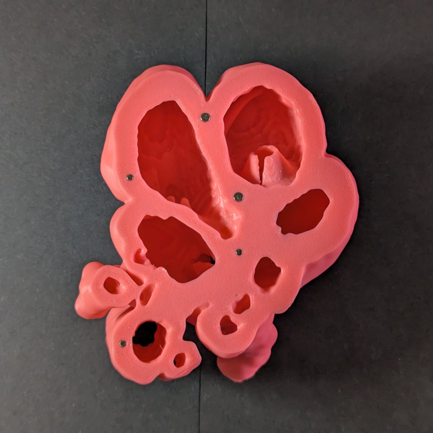
Right ventricular outflow tract and three-vessel view (RVOT/3VV)
Further up, the pulmonary artery leaves the right ventricle and crosses in front of the aorta. The RVOT view follows it out of the right ventricle to where it divides into the right and left pulmonary arteries. At the same level, the three-vessel view shows three vessels in a line: the pulmonary artery, the aorta and the superior vena cava, with the descending aorta behind them.
What you check:
- the pulmonary artery arises from the right ventricle, the aorta from the left, and the two cross rather than running side by side
- in the three-vessel view, the vessels sit in a straight or gently curved line, with the pulmonary artery on the left and furthest forward, then the aorta, then the superior vena cava on the right
- their sizes fall in the same order: pulmonary artery largest, superior vena cava smallest
Great arteries that run parallel instead of crossing are the sign to look for in transposition of the great arteries, where the four-chamber view can look normal.
Three-vessel and trachea view (3VT)
The 3VT view is the highest on the sweep, just above the three-vessel view. Here the transverse aortic arch and the ductal arch both run back towards the spine and meet the descending aorta, making a V shape. ISUOG lists the superior vena cava, main pulmonary artery, arterial duct, aortic arch and trachea in it.
What you check:
- the aortic arch and the ductal arch both lie to the left of the trachea
- the two arches are roughly the same size and, on colour Doppler, flow in the same direction
- the superior vena cava lies to the right of the aortic arch, with the trachea behind it
This is the view for arch problems. An aortic arch clearly narrower than the duct raises the question of coarctation, and an arch that passes to the right of the trachea is a right aortic arch.
How close together the views are
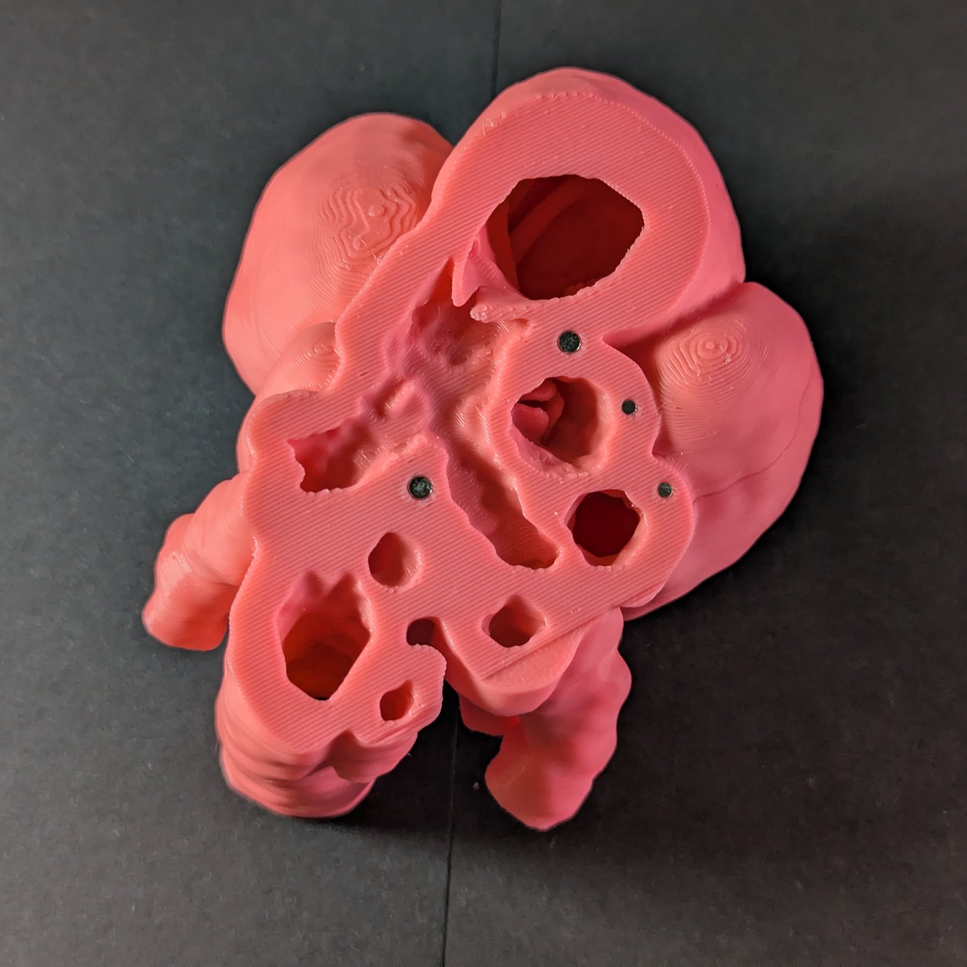
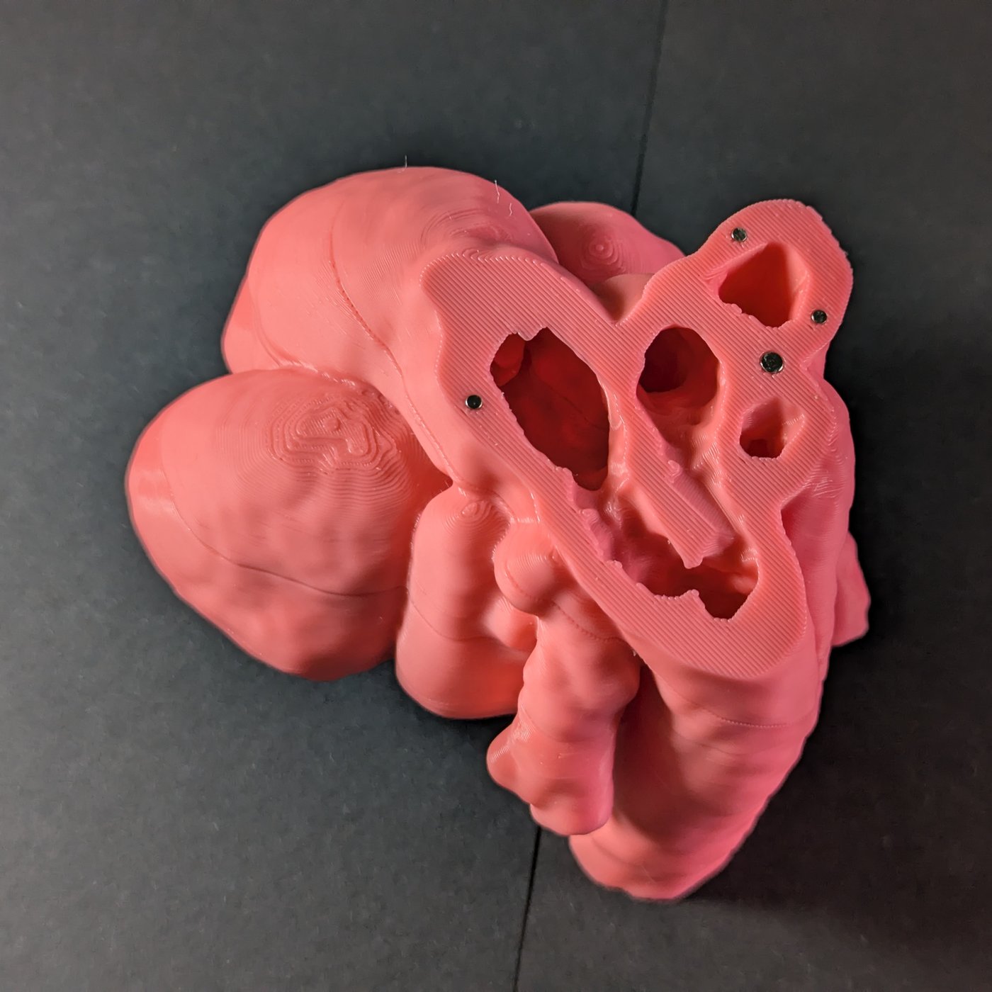
On screen each view fills the frame, so they can feel like four separate pictures. In the heart they are close together. On the life-size model, whose cardiac circumference matches a 34-week heart, the three-vessel and trachea plane is about 17 mm from the four-chamber plane, measured through the middle of the heart. The four-chamber and LVOT planes are about 7 mm apart. At the mid-trimester anomaly scan the heart is smaller, so the distances are smaller still.
That is why a slight change in probe position, or a fetus that shifts a little, takes you from one view to the next.
Learning the views on a fetal heart model
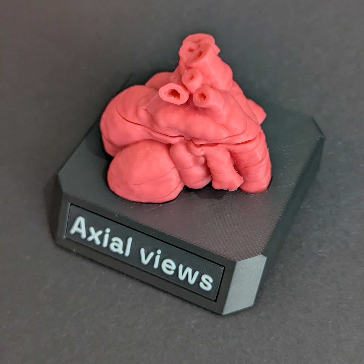
Ultrasound gives you one thin plane at a time and leaves you to build the heart in your head. A sectioned model gives you the same planes as solid pieces. Each cut face is a view you already read on screen, so you can hold the four-chamber face in one hand and the LVOT face in the other, then press the sections back together and see where each view sits in the whole heart. Each piece between two faces is the stretch of heart the probe passes through from one view to the next.
Our models are cut along these four diagnostic planes and come apart into five sections, held together by magnets and stacked in the order of the sweep, with the four-chamber view at the bottom and the 3VT at the top. The geometry comes from the King's College London fetal heart atlas, which combines the MRI of 28 healthy fetuses at 29 to 34 weeks into one averaged heart, so the anatomy is typical rather than any one patient's. How that atlas becomes a printed heart is in how we make a 3D-printed fetal heart model.
The life-size model suits one-to-one teaching or a small group around a desk. The twice-life-size model is the one for teaching a group, as its cut faces read from across a table.